Anatomy and physiology class might be inextricably linked with frogs and the odor of formaldehyde. But now, students at Wor-Wic are reaping the benefit of a more accurate, and scent-free, technology, in the form of Anatomage tables.
The high-tech tool stores digitized images of real donors, allowing students to virtually dissect human bodies. A close-up view of organs and tissues is at their fingertips, and the images can be rotated, angled and repositioned for thorough study.
“It is very helpful,” said student Carlisa Smith, “and it’s easy to use. I have never seen anything like it before.”
The tables provide real models to study closely — an invaluable tool for students, many of whom are working on degrees and certifications that will take them into patient care, such as nursing. Students in the radiologic technology program use one of the tables to learn how to position patients for medical imaging. More than just a simulation, the table contains a library of high-definition images from bodies that were donated to science.
Students can peel back layers and explore anatomical structure via the interactive, life-sized touch screen.
“My students enjoy using it,” said Dr. Subrata Biswas, assistant professor of biological science. “It is very detailed and accurate — the next best thing to a human body.”
Biswas said there are four different bodies in the virtual table, which allows students to see anatomical differences in men and women, and some of the natural variances from person to person, providing a much more in-depth experience. “It is a powerful tool for students at Wor-Wic,” he said.
Anatomy and physiology class.
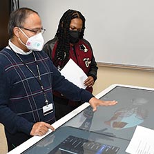
Photo: Dr. Subrata Biswas uses an Anatomage table to show lung anatomy to students in a biology class.